
Researchers have used a deep learning artificial intelligence model to discover what they describe as the primary biomarker of chronic stress that might be directly seen on standard medical images. The findings are being presented next week on the annual meeting of the Radiological Society of North America (RSNA).
Chronic stress does not only affect mood. It may possibly influence each physical and mental health, contributing to problems equivalent to anxiety, trouble sleeping, muscle pain, hypertension and a less effective immune system, in response to the American Psychological Association. Studies have also linked ongoing stress to major conditions including heart disease, depression and obesity.
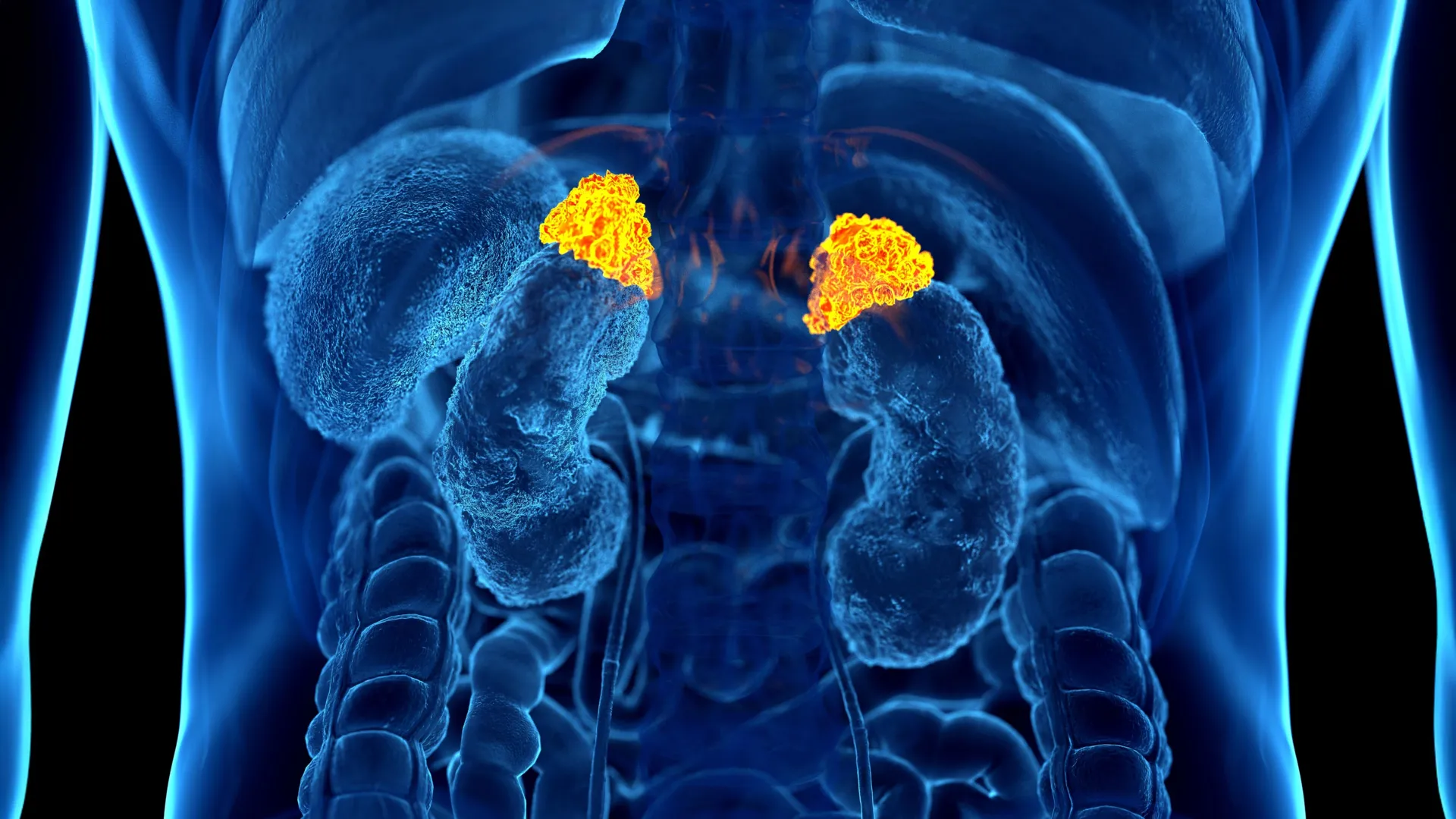
AI measures adrenal glands on routine CT scans
The study’s lead writer, Elena Ghotbi, M.D., a postdoctoral research fellow at Johns Hopkins University School of Medicine in Baltimore, Maryland, created and trained a deep learning tool designed to calculate the dimensions of the adrenal glands using CT scans that had already been performed.
Every year, tens of thousands and thousands of chest CT scans are performed in america alone.
“Our approach leverages widely available imaging data and opens the door to large-scale evaluations of the biological impact of chronic stress across a variety of conditions using existing chest CT scans,” Dr. Ghotbi said. “This AI-driven biomarker has the potential to boost cardiovascular risk stratification and guide preventive care without additional testing or radiation.”
Making the burden of stress visible within the body
Senior writer Shadpour Demehri, M.D., professor of radiology at Johns Hopkins, noted that chronic stress is amazingly common and is something many adults experience each day.
“For the primary time, we will ‘see’ the long-term burden of stress contained in the body, using a scan that patients already get each day in hospitals across the country. Until now, we’ve not had a approach to measure and quantify the cumulative effects of chronic stress, aside from questionnaires, surrogate serum markers like chronic inflammation, and cortisol measurement, which could be very cumbersome to acquire.” Dr. Demehri said.
Unlike a single cortisol test, which reflects stress at only one cut-off date, the dimensions of the adrenal glands functions more like a long-term gauge of chronic stress.
Large multi-ethnic cohort links imaging, hormones and stress load
On this research, the team analyzed information from 2,842 participants (mean age 69.3; 51% women) enrolled within the Multi-Ethnic Study of Atherosclerosis, a big study that mixes chest CT imaging, validated stress questionnaires, cortisol measurements and indicators of allostatic load — the cumulative physiological and psychological effects of chronic stress on the body. Since it integrates imaging, biochemical data and psychosocial assessments in the identical individuals, this cohort was uniquely suited, and sure the just one available, for creating an imaging-based marker of chronic stress.
The investigators applied their deep learning model to the CT scans to mechanically outline and measure adrenal gland volume. They defined Adrenal Volume Index (AVI) as adrenal volume (cm3) divided by height2 (m2). To capture hormonal patterns, participants provided salivary cortisol eight times per day over the course of two days. Allostatic load was calculated using body mass index, creatinine, hemoglobin, albumin, glucose, white blood count, heart rate and blood pressure.
Adrenal Volume Index tracks stress, hormones and heart risk
The team then examined how AVI related to cortisol, allostatic load and a variety of psychosocial stress indicators, equivalent to depression scores and perceived stress questionnaires. They found that AVI generated by the AI model aligned with established stress questionnaires, with circulating cortisol levels and with future adversarial cardiovascular events.
Higher AVI values were linked with greater overall cortisol exposure, higher peak cortisol levels and increased allostatic load. Individuals who reported high levels of perceived stress had higher AVI compared with those that reported low stress. AVI was also connected to the next left ventricular mass index, a measure related to heart structure. For each 1 cm3/m2 increase in AVI, the chance of heart failure and death increased.
“With as much as 10-year follow-up data on our participants, we were in a position to correlate AI-derived AVI with clinically meaningful and relevant outcomes,” Dr. Ghotbi said. “That is the very first imaging marker of chronic stress that has been validated and shown to have an independent impact on a cardiovascular final result, namely, heart failure.”
A brand new approach to quantify the cumulative impact of stress
“For over three many years, we have known that chronic stress can wear down the body across multiple systems,” said Teresa E. Seeman, Ph.D., study co-author and professor of epidemiology at UCLA and a pioneering researcher in stress and health. “What makes this work so exciting is that it links a routinely obtained imaging feature, adrenal volume, with validated biological and psychological measures of stress and shows that it independently predicts a significant clinical final result. It is a true step forward in operationalizing the cumulative impact of stress on health.”
Dr. Demehri explained that connecting an easy imaging measure with several well-established markers of stress and disease outcomes creates a brand new, practical approach to measuring chronic stress in on a regular basis clinical practice.
“The important thing significance of this work is that this biomarker is obtainable from CTs which might be performed widely in United States for various reasons,” Dr. Demehri said. “Secondly, it’s a physiologically sound measure of adrenal volume, which is a component of the chronic stress physiologic cascade.”
The researchers noted that this imaging biomarker could potentially be applied to many stress-related diseases that commonly affect middle-aged and older adults.
Other co-authors are Roham Hadidchi, Seyedhouman Seyedekrami, Quincy A. Hathaway, M.D., Ph.D., Michael Bancks, Nikhil Subhas, Matthew J. Budoff, M.D., David A. Bluemke, M.D., Ph.D., R. Graham Barr and Joao A.C. Lima, M.D.