
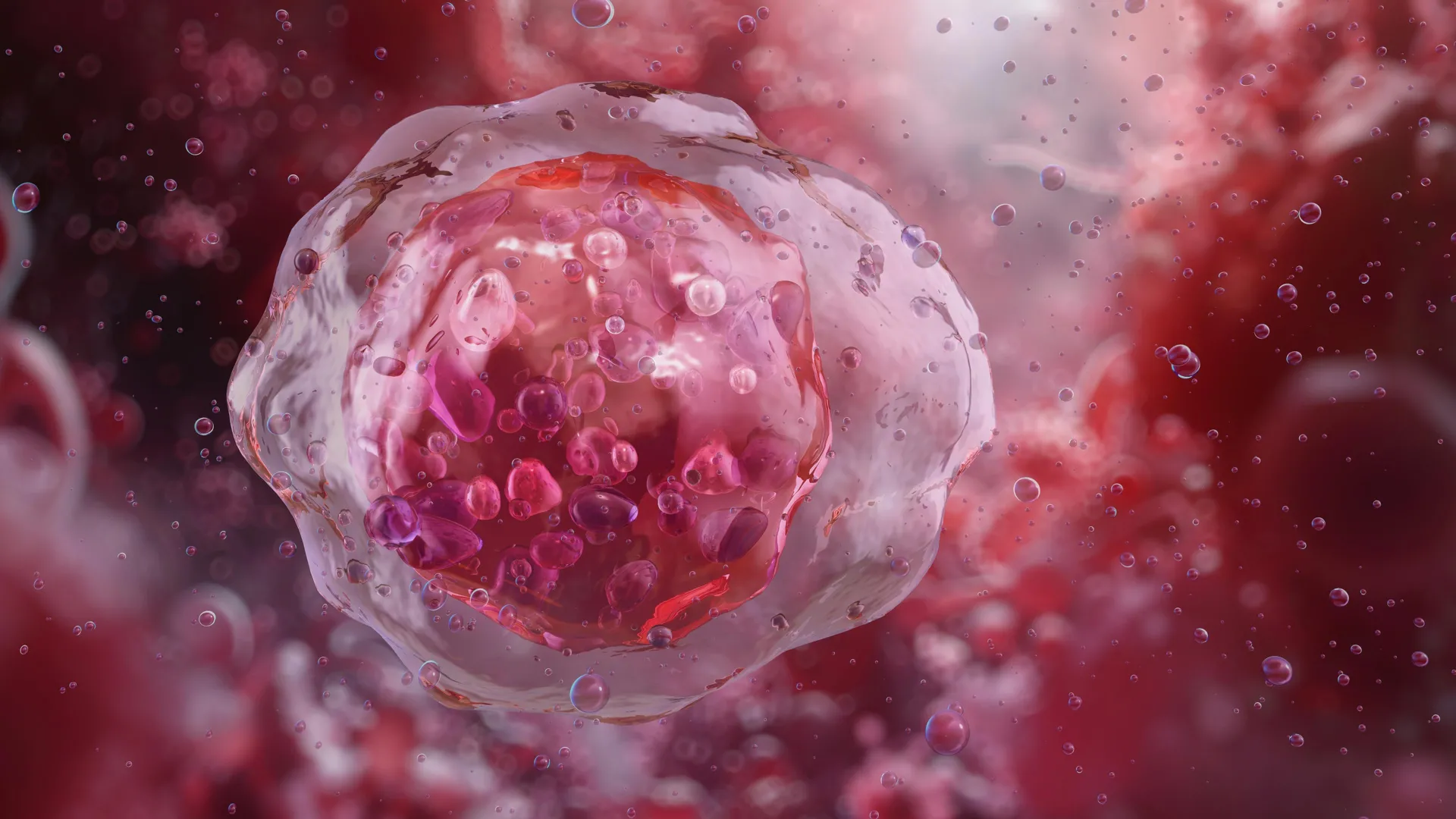
A brand new artificial intelligence system that examines the form and structure of blood cells could significantly improve how diseases resembling leukemia are diagnosed. Researchers say the tool can discover abnormal cells with greater accuracy and consistency than human specialists, potentially reducing missed or uncertain diagnoses.
The system, often known as CytoDiffusion, relies on generative AI, the identical style of technology utilized in image generators resembling DALL-E, to research blood cell appearance intimately. Reasonably than focusing only on obvious patterns, it studies subtle variations in how cells look under a microscope.
Moving Beyond Pattern Recognition
Many existing medical AI tools are trained to sort images into predefined categories. In contrast, the team behind CytoDiffusion demonstrated that their approach can recognize the total range of normal blood cell appearances and reliably flag rare or unusual cells that will signal disease. The work was led by researchers from the University of Cambridge, University College London, and Queen Mary University of London, and the findings were published in Nature Machine Intelligence.
Identifying small differences in blood cell size, shape, and structure is central to diagnosing many blood disorders. Nonetheless, learning to do that well can take years of experience, and even highly trained doctors may disagree when reviewing complex cases.
“We have all got many differing types of blood cells which have different properties and different roles inside our body,” said Simon Deltadahl from Cambridge’s Department of Applied Mathematics and Theoretical Physics, the study’s first creator. “White blood cells focus on fighting infection, for instance. But knowing what an unusual or diseased blood cell looks like under a microscope is a crucial a part of diagnosing many diseases.”
Handling the Scale of Blood Evaluation
A typical blood smear can contain 1000’s of individual cells, excess of an individual can realistically examine one after the other. “Humans cannot have a look at all of the cells in a smear — it’s just impossible,” Deltadahl said. “Our model can automate that process, triage the routine cases, and highlight anything unusual for human review.”
This challenge is familiar to clinicians. “The clinical challenge I faced as a junior hematology doctor was that after a day of labor, I’d face lots of blood movies to research,” said co-senior creator Dr. Suthesh Sivapalaratnam from Queen Mary University of London. “As I used to be analyzing them within the late hours, I became convinced AI would do a greater job than me.”
Training on an Unprecedented Dataset
To construct CytoDiffusion, the researchers trained it on greater than half 1,000,000 blood smear images collected at Addenbrooke’s Hospital in Cambridge. The dataset, described as the biggest of its kind, includes common blood cell types, rare examples, and features that usually confuse automated systems.
As a substitute of simply learning the way to separate cells into fixed categories, the AI models your complete range of how blood cells can appear. This makes it more resilient to differences between hospitals, microscopes, and marking techniques, while also improving its ability to detect rare or abnormal cells.
Detecting Leukemia With Greater Confidence
When tested, CytoDiffusion identified abnormal cells related to leukemia with much higher sensitivity than existing systems. It also performed in addition to or higher than current leading models, even when trained with far fewer examples, and was capable of quantify how confident it was in its own predictions.
“After we tested its accuracy, the system was barely higher than humans,” said Deltadahl. “But where it really stood out was in knowing when it was uncertain. Our model would never say it was certain after which be flawed, but that’s something that humans sometimes do.”
Co-senior creator Professor Michael Roberts from Cambridge’s Department of Applied Mathematics and Theoretical Physics said the system was evaluated against real-world challenges faced by medical AI. “We evaluated our method against most of the challenges seen in real-world AI, resembling never-before-seen images, images captured by different machines and the degree of uncertainty within the labels,” he said. “This framework gives a multi-faceted view of model performance which we imagine will probably be helpful to researchers.”
When AI Images Idiot Human Experts
The team also found that CytoDiffusion can generate synthetic images of blood cells that look indistinguishable from real ones. In a ‘Turing test’ involving ten experienced hematologists, the specialists were no higher than random probability at telling real images aside from those created by the AI.
“That actually surprised me,” Deltadahl said. “These are individuals who stare at blood cells all day, and even they couldn’t tell.”
Opening Data to the Global Research Community
As a part of the project, the researchers are releasing what they describe because the world’s largest publicly available collection of peripheral blood smear images, totaling greater than half 1,000,000 samples.
“By making this resource open, we hope to empower researchers worldwide to construct and test latest AI models, democratize access to high-quality medical data, and ultimately contribute to higher patient care,” Deltadahl said.
Supporting, Not Replacing, Clinicians
Despite the strong results, the researchers emphasize that CytoDiffusion shouldn’t be intended to exchange trained doctors. As a substitute, it’s designed to help clinicians by quickly flagging concerning cases and mechanically processing routine samples.
“The true value of healthcare AI lies not in approximating human expertise at lower cost, but in enabling greater diagnostic, prognostic, and prescriptive power than either experts or easy statistical models can achieve,” said co-senior creator Professor Parashkev Nachev from UCL. “Our work suggests that generative AI will probably be central to this mission, transforming not only the fidelity of clinical support systems but their insight into the boundaries of their very own knowledge. This ‘metacognitive’ awareness — knowing what one doesn’t know — is critical to clinical decision-making, and here we show machines could also be higher at it than we’re.”
The team notes that additional research is required to extend the system’s speed and to validate its performance across more diverse patient populations to make sure accuracy and fairness.
The research received support from the Trinity Challenge, Wellcome, the British Heart Foundation, Cambridge University Hospitals NHS Trust, Barts Health NHS Trust, the NIHR Cambridge Biomedical Research Centre, NIHR UCLH Biomedical Research Centre, and NHS Blood and Transplant. The work was carried out by the Imaging working group throughout the BloodCounts! consortium, which goals to enhance blood diagnostics worldwide using AI. Simon Deltadahl is a Member of Lucy Cavendish College, Cambridge.